Small anatomical reminder
It is located to the right of the epigastric hollow, in the right hypochondrium. It is the largest of the viscera with a weight of 1500 grams. It is surrounded by a capsule called the Glisson capsule.
It consists of 2 lobes: the right lobe which represents 2/3 of the liver and the left lobe separated by the suspensory ligament.
The liver is divided into sectors by the suprahepatic veins, which are themselves divided into 8 segments by the portal vein.
Oxygenated blood is supplied by the hepatic artery and venous blood, rich in all nutrients, is supplied by the portal vein of the esophagus, stomach and intestines. These 2 main vessels are divided into different arteries and veins that irrigate the liver. The supra-hepatic veins ensure the venous return of the liver to the inferior vena cava.
The functions of the liver are multiple:
• Nutritional function: it is involved in the metabolism of carbohydrates and fats and in the regulation of blood sugar levels.
• Synthesis function: of proteins, bile, bile acids (which help digest fat)
• Endocrine function: Synthesis of different hormones and vitamin D
• Detoxifying function: eliminates toxins, drugs and other waste from the body
• Storage function: vitamins B2, A, D, D, E, K, iron, copper, sugar, which it releases when the body needs it.
The body only needs 1/4 of the liver to live and the liver has the extraordinary function of being able to regenerate itself.
LIVER TUMORS:
A tumor is when a mass is discovered. Not all tumours are therefore cancers such as angiomas, cysts, adenomas and focal nodular hyperplasias, for example, which do not systematically require surgical intervention.
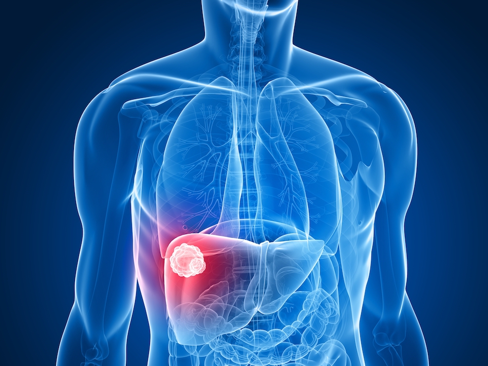
HEPATOCELLULAR CARCINOMA
The liver is mainly composed of lobular hepatocytes, which are responsible for 90% of primary liver cancers called hepatocellular carcinoma. Other, rarer tumours exist from other cells that make up the liver.
Liver metastases are completely different. They correspond to cancer cells from other organs that invade the liver. The treatment is different and depends on the cancer of origin.
EPIDEMIOLOGY:
This cancer has been on the rise for several decades. There are an average of 8500 new cases per year. It is the 7th cancer in France. It affects men more than women. Its average age of diagnosis is 63 years.
RISK FACTORS:
• Tobacco
• Chronic alcoholism
• Chronic hepatitis B and C
• Liver steatosis: excess fat in the Liver often linked to obesity and diabetes
• Hemochromatosis: excess iron in the body, genetic disease
Duration of hospital stay
Variable.
The time spent abroad will depend on the treatment.
Average length of stay
Long stays.
Several long stays are sometimes necessary.
Every year, nearly 11 million patients go abroad in search of medical care. At MEDICAIM, we provide our patients with access to the best hospitals and doctors around the world. Contact us to learn more about your treatment options.
Ask for your free quote abroad
Start your medical stay by requesting a quote. Our customer service department will help you find the clinic that best suits your needs and get you a quote.
THE DIAGNOSIS:
The abdominal Doppler ultrasound:
It is done quickly by placing a probe on the belly that emits ultrasonic waves through a gel. These waves make it possible to see the organs of the abdomen that are visualized on a screen and the vessels with their blood flows evaluated by Doppler.
Ultrasound is often the initial examination that suggests cancer: large liver, nodules, dilation of vessels, portal flow inversion, large spleen.
Blood dosage:
• Standard dosage which includes the liver test (transaminases, gamma GT, alkaline phosphatases) which is disrupted (increased) in case of cancer.
• Determination of specific tumor markers: Alphafoetoprotein is increased, Carcino-embryonic Antigen (CEA) CA19-9 are normal.
• Determination of Liver proteins (prothrombin and albumin) to assess Liver function.
**THE THORACO-ABDOMINO-PELVIC CT: **
It is the essential examination to make the etiological diagnosis and look for metastases and damage to blood vessels. It is made with and without contrast injection.
**THE HEPATIC MRI (MAGNETIC RESONANCE IMAGING): **
It is carried out with Gadolinium injection. Allows to perfectly specify the tumor and performed in addition to or in replacement of the scanner.
ESOGASTRODUODENAL FIBROSCOPY:
It is performed under local or general anesthesia. Consists of passing a tube with a camera through the mouth to the stomach to assess the potential presence of esophageal varices. Biopsies can be performed on the trajectory.
HEPATIC BIOPSY:
It is performed under general anesthesia under ultrasound control. It provides histological evidence of the diagnosis of hepatocellular carcinoma and thus eliminates a metastasis of another cancer.
The choice of treatment is made during a multidisciplinary meeting.
SURGERY:
This is a partial removal of the liver. This procedure can be performed if the cancer is small (3 to 5 cm) in a non-cirrhotic liver or in a cirrhotic liver with moderate hepatic failure. It is totally contraindicated in case of portal hypertension (too high pressure in the vessels).
A consultation is first carried out with the surgeon and the anaesthetist. The surgeon asks you to sign an informed consent form so that you can authorize him or her to remove a piece of the tumour in order to keep it for future research.
The intervention:
The anaesthetist places a nasogastric tube through the nose and into the stomach. It allows the digestive system to rest and avoids post-operative complications. A urinary catheter is also placed.
Surgery is usually performed by laparotomy: opening of the abdomen. Sometimes it is performed by laparoscopy (4 incisions are made and the surgeon puts his/her material under screen).
In all cases, ultrasound monitoring is performed during the procedure to locate the cancerous nodule and the surrounding blood vessels.
The surgeon removes the segment containing the nodule and may also have to remove the segments around it. This is called segmentectomy. When it removes an entire lobe, it is called a lobectomy.
When the liver is too damaged by cirrhosis, the surgeon is forced to remove only the tumor. Regardless of the type of procedure, the surgeon systematically removes a healthy part surrounding the tumor called the resection margin.
Drains are placed in the operated area to evacuate blood and lymph and avoid swelling and hematoma.
The cancerous part is sent to the anatomopathology laboratory for study.
The consequences of the intervention:
You stay in the recovery room for about 2 hours. The anaesthetist sets up an anti-pain treatment by venous route based on morphine. Injections to prevent phlebitis are introduced daily.
You are then hospitalized in intensive care for close supervision for the first 2 to 3 days. The probes are removed before you leave for the gastroenterology department.
Complications are monitored several times a day, which can be:
• Hematoma or wound infection
• Hemorrhaging
• Pain in the operated area
• Diffuse Pain
• Hepatic insufficiency.
If there are no complications, the liver regenerates quickly and a gradual recovery is achieved.
The discharge takes place between the 8th and 10th day.
Monitoring by blood alfafoetoprotein assay and hepatic ultrasound is performed at 3 months and then every 6 months for 2 years.
HEPATIC TRANSPLANTATION or LIVER TRANSPLANT:
It treats both hepatocellular carcinoma and cirrhosis.
Due to the lack of an organ, it cannot be applied to all patients.
There are also contraindications:
• Over 65 years old
• Long-term drug treatment incompatible with anti-rejection treatment
• Chronic unweaned alcoholism
• Metastasis and invasion of blood vessels.
• Size and number of tumours (>5cm or >3 tumours of >2 cm).
Liver transplantation requires an additional check-up.
TISSUE REMOVAL BY RADIOFREQUENCY:
This consists of destroying the tumor by heat.
It is performed when the surgical procedure is contraindicated.
It is performed under general anesthesia by a specialized radiologist.
Prior consultation with the radiologist and anesthesiologist is required.
The intervention:
The radiologist passes a thin needle through the abdomen facing the liver under ultrasound control. It then sends heat of the order of 50°C for 6 to 12 minutes. He/she then removes the probe still held at 50°C to kill the remaining potential cancer cells. The destruction of the tumor is controlled by ultrasound.
In the recovery room, pain treatments have begun. The hospitalization lasts 24 to 48 hours.
Potential complications are: infection or hematoma at the injection site, pain.
A control with an MRI at 2 months is carried out and then every 6 months for 2 years.
ARTERIAL LIPIOEMBOLIZATION CHEMO:
This treatment is indicated when surgery is not possible or awaiting transplantation. It is impossible in case of damage to the portal vein. This involves combining chemotherapy (drug treatment that kills cancer cells), lipiodol and embolization (blocking the tumor's blood vessels).
The type of chemotherapy depends on the tumour. Before any intervention, a specialized appointment is made with the radiologist who carries out the examination and the anaesthetist. A pre-intervention MRI is scheduled.
The intervention takes place in a specific room. The radiologist passes a catheter through the femoral artery and brings it to the liver under radio control and injects iodized product. He then sends the products that embolize the vessels (block the blood and kill the cancer cells) and the chemotherapy drug.
Hospitalization lasts 24 to 48 hours.
Side effects are monitored.
For chemotherapy, the side effects can be: hair loss, nausea, fatigue.
For embolization complications can be: fever, abdominal pain
TARGETED THERAPY:
This consists of giving a drug that specifically kills cancer cells. The only current treatment is Sorafenib (NEXAVAR). 800mg/day is prescribed in 2 doses.
A pre-intervention assessment and follow-up are necessary.
Contraindications: esophageal varices and high blood pressure.
Some complex diseases such as cancer can lead some patients to seek a second medical opinion. Almost 50% of patients using the second medical opinion have seen their treatment options evolve. Seeking a second medical opinion is perfectly legitimate when faced with a serious illness.
Click here to find out more about the second MEDICAIM medical opinion
MEDICAIM takes care of the follow-up on a case-by-case basis. www.medicaim.com
MEDICAIM is looking for the best specialists for you and we will offer you several renowned doctors.
MEDICAIM organizes your entire stay for you: post-operative nursing care, biological follow-up, therapeutic, nutritional and psychological support.
Any additional questions? Ask your MEDICAIM doctor about it: careteam@medicaim.com
Some needs and conditions are more complex than others. In case of doubt, please send us additional information to establish a customized quote.
Ask for a quoteCertains besoins et pathologies sont plus complexes que d’autres. En cas de doute, faîtes-nous parvenir des informations complémentaires pour établir un devis sur-mesure.
Demander un devisEntrust us with your medical file and it will be examined by a specialist doctor. The goal?
Allow you to evaluate all your treatment options.