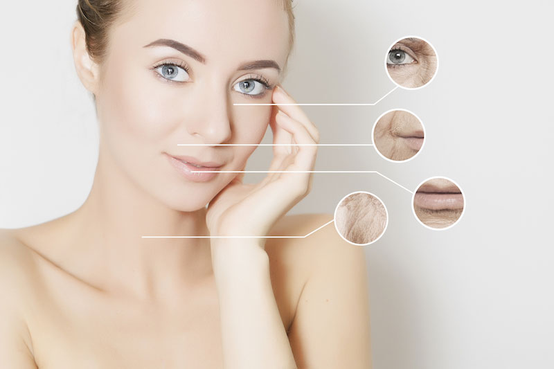
Skin tumors (or lesions) are spots or growths of the skin of varying size, shape and colour. They can be located on the entire face or body.
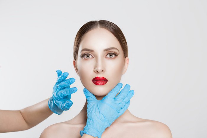
Each cell type in the skin is capable of turning into a benign or malignant tumor, so there are a large number of skin tumors, ranging from a simple "mole" to a very rare tumor. They can appear during life or be present from birth. They are most often diagnosed by a dermatologist or by your attending physician who asks a plastic surgeon to perform the removal.
With regard to skin tumors, three different cases can be distinguished:
** Benign tumors:**
Among benign tumours, are most frequently found:
Melanoma tumors:
These are extremely frequent injuries. There are different forms such as epheleids, also called "freckles", which are related to sun exposure. Lentigos are almost similar except that they are present all year round.
Moles can be present at birth or appear throughout life. They can affect all parts of the body.
Epidermal tumors:
The most frequently found are seborrheic warts (or seborrheic keratoses). They often appear after forty years. They have a warty appearance and are brown and more or less pigmented.
Keratoses (or actinic keratoses) are small brown or red spots located in areas exposed to the sun.
Some viral infections can affect the epidermis, such as viral papillomas responsible for the appearance of warts (flat, vulgar, plantar), condylomas (ano-genital) or molluscum contagiosum.
Adnexal tumors:
These are lesions developed at the expense of hair cells. The most frequently found are cysts. There are several cyst entities such as sebaceous cysts, epidermal cysts and milium granules of the face or trichilemnaux cysts.
There are other lesions but they occur even rarer: keratoacanthoma, pilomatricoma tricho-epithelioma…
Tumors of connective tissue:
They develop from the connective tissue, vessels, nerves, fat, muscles contained in the skin. There are again many of them.
The main ones are histiocyto bromes and molluscum pendulum.
Vascular lesions include angiomas. Botriomycoma is a very frequent conjunctival vascular lesion whose starting point is often a small inflammatory wound.
Lipomas are very common fat tumours. They can be unique or multiple.
Neuro bromes are nerve tumours; when they are multiple, they must be suggestive of Recklinghausen disease.
Xanthelasmas are lipid deposits often found on the eyelids.
Questionable tumors: the simple visual examination of a lesion does not always make it possible to affirm its benign or malignant nature.
All the benign tumors mentioned above can be included in this category if their appearance, shape or situation is unusual. Melanoma tumours are particularly important in this category, especially when they are very large and present from birth or if they are traumatized, if their colour or border changes if they thicken.
They can turn or cause a diagnostic problem with malignant melanoma. Similarly, it is recommended to remove them if their anatomical situation may lead to a risk of trauma or pose a surveillance problem (plantar region, hand, back, oral or genital mucosa, conjunctiva, scalp).
Malignant tumors: skin cancers must of course be removed because surgery is often the only treatment that can achieve a complete cure. The aim is therefore to remove them completely by allowing a "safety margin", i.e. by passing offshore on the sides and at depth in order to give yourself every chance to avoid a recurrence.
There are three main categories of malignant skin tumors:
Epitheliomas: these are extremely frequent lesions. There are different forms such as basal cell epitheliomas and spino-cellular epitheliomas also called basal cell carcinomas and spino-cellular carcinomas.
Basal cell epitheliomas are the most common cancers. They generally occur in white-skinned people on average after 45 years of age. Their main risk factor is sun exposure. They affect the face and neck in more than 80% of cases. Their evolution is purely local without metastasis, but a neglected evolution can lead to significant mutilations, especially when the tumor is located near the eyes, nose, mouth or ears. Usually the surgeon removes the lesion with a safety margin of a few millimeters.
Squamous cell epitheliomas are also called squamous cell carcinomas. They usually occur after the age of 40. Their main risk factor is sun exposure. They appear mainly on areas exposed to the sun (face, hands) and also affect the mucous membranes (buccal, anal or genital). In some cases there is a risk of lymph node involvement and therefore metastasis.
Melanomas: Malignant melanoma is much less frequent than previous tumors. It most often occurs on fair skin and sun exposure is a major risk factor. It may take the form of a recently occurring black pigmented lesion on healthy skin. It can also occur on a pre-existing "mole". All parts of the body can be affected, including the mucous membranes and conjunctiva. When melanoma is confirmed, treatment is essentially surgical and consists of removing the lesion with fairly wide safety margins. The search for lymph node involvement will be systematic. Depending on the initial thickness of the melanoma and the lymph node status, additional treatment may be offered (lymph node cleaning, chemotherapy, immunotherapy).
Sarcomas: these are rare tumors. The anatomical presentations and locations are diverse and varied. The treatment is primarily surgical. Radiotherapy and/or chemotherapy may be indicated in addition to the surgical procedure.
Duration of hospital stay
1 day.
Pure local anaesthesia.
Average length of stay
1 to 2 days.
If classic general anesthesia during which you sleep completely."
Every year, nearly 11 million patients go abroad in search of medical care. At MEDICAIM, we provide our patients with access to the best hospitals and doctors around the world. Contact us to learn more about your treatment options.
Ask for your free quote abroad
Start your medical stay by requesting a quote. Our customer service department will help you find the clinic that best suits your needs and get you a quote.
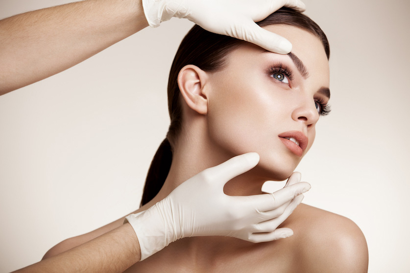
An interview followed by an examination of the skin lesion will have been carried out by the surgeon in order to specify the surgical possibility(s).
In the event of anaesthesia other than purely "local" anaesthesia, a pre-anaesthetic check-up may be prescribed and a pre-operative consultation with the anaesthetist is mandatory.
No medication containing aspirin should be taken within 10 days of the procedure.
Depending on the type of anesthesia, you may be asked to fast (no food or drink) 6 to 7 hours before the procedure.
No make-up, jewellery or piercing should be worn during the operation.
**Type of anesthesia: Three procedures: **
Pure local anaesthesia where an analgesic product is injected to ensure the insensitivity of the area to be operated on. This is the most frequent case for basic Dermato-Surgery.
Vigil anaesthesia (local anaesthesia deepened by tranquilizers) during which you can stay awake but where you will be relaxed and from which some amnesia about the operation may result. It may be preferred for reasons of personal comfort or for the realization of certain complex flaws, especially on the face.
Classical general anaesthesia during which you sleep completely, in fact rarely useful in Dermatology and Surgery.
The choice between these different techniques will be the result of a discussion between you, the surgeon and the anesthesiologist.
Hospitalization conditions:
Basic Dermato-surgery procedures, especially if they are performed under pure local anaesthesia, do not necessarily require hospitalisation and can, like dental treatment, be carried out in an office, provided all the necessary equipment is available.
If the procedure is scheduled in a clinic or hospital, it can usually be performed " externally ", i.e. with an entry just before the operation and an exit just after the operation or " ambulatory ", i.e. in "day hospitalisation" with a discharge on the same day after a few hours monitoring. Traditional hospitalization with a night to spend on site is exceptional for this type of surgery.
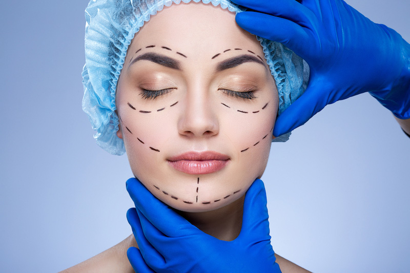
The removal of a skin tumor, whether small or large, remains above all a surgical procedure that requires the respect of certain principles.
As with any surgery, it must be performed in good technical (suitable rooms, lighting, instruments) and hygienic conditions (asepsis of the patient, washing the surgeon's hands, wearing sterile gloves, mask…).
Surgical exeresis must remove the lesion completely with often healthy skin safety margins. A histological analysis of the lesion is systematic because only this examination can confirm the benign or malignant nature of the lesion. In some cases, this analysis makes it possible to assess the aggressiveness of the tumor. In the case of a large or poorly located lesion or if the malignant nature is not certain, your surgeon may need to perform a biopsy to identify the lesion. The surgical technique and reconstruction procedures may be different depending on the type of lesion.
Depending on the size and location of the tumor, there are several methods of repairing the loss of substance.
The only aggressions inflicted on the skin that disappear without leaving any scars are those that only concern the most superficial part of the skin, namely the epidermis.
As soon as an incision crosses the dermis, i. e. the deep part of the skin, and whatever the quality of the surgeon and the care provided by him/her, the surgical procedure will leave behind a scar which, of course, will diminish and become more or less discreet, but never totally invisible.
In the case of skin tumors, the aim is to remove the lesion completely, with a safety margin for malignant lesions, the extent of which depends on the type of lesion, and to achieve as discreet a scar as possible.
The basic principle is "spindle" ablation followed by direct suturing by moving the banks closer together. The integration of the area to be removed into a spindle is essential to avoid the formation of folds at the ends of the scar during closure but results in a scar whose size is larger than the diameter of the initial lesion. On this subject, it should be noted that if the scar is often larger than the initial lesion, it is because lengthening a scar reduces the tension that is exerted on each of its edges and thus to have the best possible aesthetic result in the long term.
In addition, the discretion of the scar will be enhanced by the orientation of the incision in line with the natural folds of the skin and by an irreproachable suture technique.
In cases where the size of the lesion or its location makes direct suture closure impracticable, the coverage of the removed area will be provided either by a skin graft taken from another region or by a local plasty corresponding to the displacement of a neighbouring skin flap so that it covers the loss of skin substance. The scarring cost of this type of flap is of course higher, but the aesthetic results are often better than those of a graft.
You may experience some discomfort with a feeling of tension on the scar, but real disabling pain is rare.
During the first few days, it is important to avoid "forcing" the scar. Caution should be exercised with regard to movements that would place too much stress on the operating area.
In the hours following the procedure, a small oozing of blood (red) or lymph (yellow) may slightly stain the bandage. Within the first 48 hours, the operated area may also sometimes show edema (swelling) and small bruises that are only transient.
Itching is also quite frequent during the healing phase. All these findings are not worrying and should be considered as "usual" consequences.
Stitches, when they are not absorbable, are removed between the 5th and 15th day.
The scar can then be massaged according to your surgeon's instructions.
Concerning exposure to the sun, as long as the scar is still dark, it is preferable to avoid any exposure and to use a "total sunscreen" type of protection.
A period of several months (sometimes up to one to two years) is necessary to assess the final appearance of the scar.
It must be understood that scarring remains a random phenomenon whose quality can in no way be guaranteed. The perfect technical expertise of a qualified plastic surgeon specifically trained for this type of operation makes it possible to put all the chances on ones side but does not eliminate this random aspect.
It is essential to regularly monitor the evolution and appearance of the scar. This is the only way to ensure that possible scarring disorders are identified in time and that appropriate treatment is applied.
MEDICAIM organizes your entire stay for you: post-operative nursing care, biological follow-up, therapeutic, nutritional and psychological support.
Any additional questions? Ask your doctor about it: careteam@medicaim.com
Some needs and conditions are more complex than others. In case of doubt, please send us additional information to establish a customized quote.
Ask for a quoteCertains besoins et pathologies sont plus complexes que d’autres. En cas de doute, faîtes-nous parvenir des informations complémentaires pour établir un devis sur-mesure.
Demander un devisEntrust us with your medical file and it will be examined by a specialist doctor. The goal?
Allow you to evaluate all your treatment options.